Digital X-ray

Clear, high‑quality images at lower radiation doses

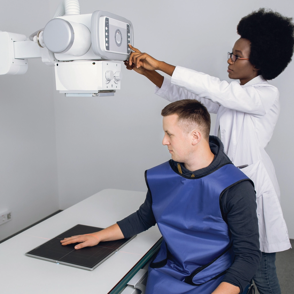
X-rays have been used in medical imaging for more than 100 years. An X-ray examination is a quick and painless imaging test that uses a small amount of radiation to create images of the inside of your body. The X-ray used at Parker Count Imaging uses the latest digital technology. It offers significant advantages over traditional film x-rays, including less radiation exposure and superior image quality that can be digitally enhanced to assist in providing an accurate diagnosis.

X-ray is often used to clearly see structures like bones and detect issues such as fractures, infections, or certain lung conditions.

Digital X-ray

How does digital x-ray work?

It works by passing small, controlled amounts of radiation through your body, where different tissues—such as bones, organs, and muscles—absorb the radiation in different ways. Dense structures like bone absorb more radiation and appear white, while softer tissues allow more rays to pass through and appear darker.

A detector on the other side captures this pattern and turns it into a detailed image that your healthcare provider can review. The process is very quick, painless, and uses only a very low dose of radiation.

What can I expect during my X-ray?

During an X-ray exam, the technologist will position you standing, sitting, or lying down, depending on the area being imaged. You may be asked to hold still or briefly hold your breath to ensure clear pictures. The equipment may be moved around you, but nothing will touch you except for positioning aids. The exam itself is painless and usually takes just a few minutes. In some cases, you may be asked to remove jewelry or wear a gown to avoid anything that could interfere with the images, and you can return to your normal activities following your exam.

For more information about digital x-ray at Parker County Imaging or to schedule an appointment please contact us.