Types of Medical Imaging Explained: A Beginner’s Guide
If your doctor just told you that you need “some imaging,” you’ve probably already opened a browser tab and typed something like Which types of medical imaging do I need? It’s a fair question, and a confusing one — there isn’t just one kind of scan. There are several, and each one is built to see something the others can’t.
This guide walks through the main types of medical imaging, what each one is actually good at, and how to think through which test fits your situation. By the end, you should also have a better sense of what to look for when searching for medical imaging near me.
Why There Are Multiple Types of Medical Imaging
Different tissues in the body behave differently depending on how they’re probed — with X-rays, sound waves, magnetic fields, or radioactive tracers. A single machine can’t do all of that well. That’s why hospitals and imaging centers offer a whole toolkit of options, and why the National Institute of Biomedical Imaging and Bioengineering (NIBIB) funds ongoing research into refining each of these tools rather than replacing them with one universal scanner.¹
Here’s a look at the major types of medical imaging, roughly in the order most people encounter them.
X-Ray
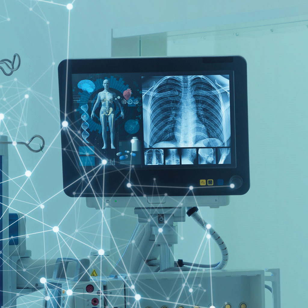
X-ray is usually the first stop for imaging, and for good reason — it’s fast, widely available, and relatively cheap. It works by passing a small amount of radiation through the body; dense tissue like bone absorbs more of it and appears white on the image, while soft tissue lets more through and appears darker. This makes X-ray the go-to for broken bones, chest infections, and dental checkups.
The tradeoff is that X-ray isn’t great at distinguishing between different types of soft tissue, so it’s rarely the final answer for anything involving organs, muscles, or ligaments.
CT Scan
A CT (computed tomography) scan is essentially a more powerful, three-dimensional version of an X-ray. Instead of a flat image, a CT scanner takes dozens or hundreds of X-ray slices and stitches them into a detailed cross-sectional picture. This makes it far better at spotting internal bleeding, tumors, and complex fractures.
Because CT relies on X-rays, it does involve more radiation exposure than a standard X-ray. According to RadiologyInfo.org, an abdomen and pelvis CT scan exposes a patient to roughly the equivalent of a few years’ worth of natural background radiation — a dose that’s generally considered acceptable when weighed against the diagnostic benefit, but one your doctor will factor in before ordering one, especially for children or during pregnancy.²
MRI
MRI (magnetic resonance imaging) uses a strong magnetic field and radio waves rather than radiation, which is why it’s often preferred for soft tissue, the brain, the spine, and joints. NIBIB explains that MRI works by aligning hydrogen protons in the body’s water molecules with a magnetic field, then measuring the energy they release as they realign after a radio pulse — different tissues release that energy at different rates, which creates contrast in the image.¹
MRI takes longer than a CT scan, often 30 to 60 minutes, and the machine is loud and enclosed, which can be uncomfortable for people with claustrophobia. But because it avoids ionizing radiation, it’s often the safer long-term choice when repeated imaging is needed.
Ultrasound
Ultrasound uses high-frequency sound waves instead of radiation or magnets, bouncing them off internal structures and interpreting the echoes to build a real-time image. It’s the imaging test most people associate with pregnancy, but it’s also used for the heart, abdominal organs, blood vessels, and guiding needle biopsies.
It’s generally the least invasive and least expensive option, with no radiation exposure, though image quality can be limited for deeper structures or patients with higher body mass.
PET Scan and Nuclear Imaging
A PET (positron emission tomography) scan takes a different approach entirely — instead of imaging structure, it images function. A small amount of a radioactive tracer is injected into the body, and the scanner tracks where it accumulates. Since cancer cells and certain other tissues metabolize the tracer differently than healthy cells, PET scans are especially useful for cancer staging, monitoring treatment response, and evaluating brain or heart function. PET is often combined with CT in a single session so doctors get both the functional and structural picture at once.
Which Imaging Test Do I Need?
This is ultimately a decision your doctor makes based on your symptoms, medical history, and what they’re trying to rule in or out — but it helps to understand the general logic they’re working from:
- Suspected broken bone or lung issue → X-ray first
- Internal bleeding, complex fracture, or a fast, detailed scan in an emergency → CT scan
- Brain, spinal cord, ligament, or soft tissue detail, without radiation → MRI
- Pregnancy monitoring, heart function, or a quick, radiation-free look at organs → Ultrasound
- Cancer staging or tracking how a disease is behaving over time → PET scan
The American Cancer Society notes that the right imaging test often comes down to balancing diagnostic benefit against radiation exposure, and that doctors weigh this tradeoff differently depending on the patient’s age and the urgency of the situation.³ That’s part of why the same symptom can lead two different doctors to order two different types of medical imaging — it’s not inconsistency, it’s judgment.
Finding Medical Imaging Near You
Once you and your doctor have settled on a test, the next step is usually logistics. A few things worth checking before you book:
- Whether the facility is accredited (hospital-based imaging centers and many freestanding clinics carry accreditation from bodies like the American College of Radiology)
- Whether your insurance requires a specific in-network provider
- Wait times — hospital imaging departments can be slower than standalone outpatient imaging centers for non-urgent scans
A quick search for types of medical imaging near me will usually surface both hospital radiology departments and independent imaging centers in your area, and it’s worth comparing both, since pricing and appointment availability can vary significantly between the two.
The Bottom Line
There’s no single “best” imaging test — only the right test for what your doctor is trying to see. X-ray and CT are built around radiation and structure; MRI trades speed for detail without radiation; ultrasound offers a fast, radiation-free option, and PET reveals function rather than just anatomy. Understanding what each one does well makes it easier to follow along with your doctor’s reasoning.
Call Parker County Imaging to learn more about the types of imaging we offer and to schedule your test.
Sources
- National Institute of Biomedical Imaging and Bioengineering (NIBIB), Magnetic Resonance Imaging (MRI). nibib.nih.gov
- RadiologyInfo.org, Radiation Dose from X-Ray and CT Exams. radiologyinfo.org
- American Cancer Society, Understanding Radiation Risk from Imaging Tests. cancer.org